Automated cell counters represent a major step forward in terms of cell counting efficiency and precision. With a choice of count settings and protocols, they can easily be integrated into almost any workflow concerning various cell types. This doesn’t necessarily mean that your new instrument will have plug-and-play functionality automatically tuned to your specific primary lines, however.
To make the most of your automated cell counter, you can optimise the default protocols to suit specific applications or cell types. This ensures maximum accuracy that far outstrips manual counts and viability tests.
Key Settings of Interest
When fine-tuning the protocols of your automated cell counter for specific samples, there are several main parameters to bear in mind.
- Diameter: The total size range of relevant cells expressed as a minimum and maximum value.
- Roundness: A sub-set of values determining the relevant cell morphology for different channels (i.e. live/dead roundness).
- Threshold: The level of intensity required for inclusion in a count.
Recommended Settings for Different Cell Types
The accuracy of your automated cell count depends on the specificity of your protocol settings. Incorrect minimum diameters may lead to cellular debris being included. Poorly tuned thresholds can lead to mischaracterisation of live/dead cells. It is important to get your initial setup right to ensure you are extracting the most value from your automated cell counter. Here we will consider the varying best settings for Chinese hamster ovary (CHO) cells as a brief case study about the importance of automated cell counter best practices.
Case Study: Chinese Hamster Ovary Cells (CHO)
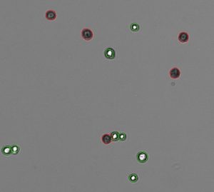
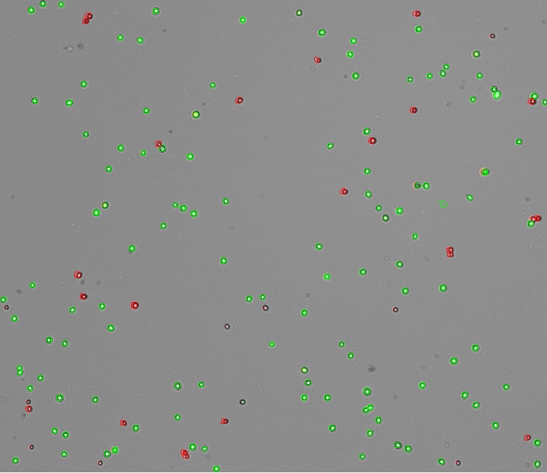
Derived from the ovary of the Chinese hamster, CHO cells are an epithelial cell line used predominantly for recombinant proteins. Counting and cell viability can measured using the fluorescent molecules acridine orange and propidium iodide, or using brightfield optics and trypan blue. Diameters vary slightly. The full, optimal range for the AO/PI App is 4—24 µm while the effect of trypan blue on cells changes means a cell diameter range of between 8—30µm is used. Live and dead roundness differ again; a value of 1 for both channels in AO/PI, and 60 (live), 25 (dead) in Trypan Blue.

As the AO/PI App uses a red and green fluorescence channel to detect both live and dead nucleated cells, you are able to set an ideal threshold for each. Both green and red thresholds ideally should be set at 1 identifying all fluorescence as originating from the cells of interest. Trypan Blue is used for brightfield viability which means you only need to establish an overall stained threshold. For best results, we recommend a stained threshold of 35 for trypan blue viability. This can be adjust to remove any cellular debris from analysis.
Automated Cell Counters for Various Cell Types
DeNovix is one of the industry’s leading suppliers of advanced instrumentation for biomedical and life sciences applications. We offer CellDropTM automated cell counters to labs with varying research tasks, including primary cell line counting and viability testing. If you would like to see a full list of recommended settings for a choice of common cell types, refer to our CellDrop Protocol Settings. Or, contact us today with any questions.


