Technical Note 187

Counting PBMCs

Introduction

It is challenging to accurately quantify and qualify the viability of primary cell samples using traditional cell counting methods. Primary cell suspensions often contain a heterogeneous collection of cell types and cellular debris as a result of digestion. Cell concentrations can also vary greatly as a result of sample location, patient or technician. Peripheral Blood Mononuclear Cells (PBMCs) are a common primary cell source used in a wide range of studies to measure immunological functions. Accurate enumeration is essential in assays such cell proliferation or cytotoxicity.

Traditional counting methods involve the use of light microscopy and hemocytometers. In addition to the inherent variability in counting between human operators, PBMCs are difficult to discern from red blood cells (RBCs). PBMCs often appear faint using light microscopy and differentiating the biconcave shape of RBCs from leukocytes requires an experienced operator and special optical setup.

The CellDrop™ FLi is an automated cell counter with high performance dual fluorescence and brightfield optics. The unique DirectPipette™ Technology eliminates disposable slides, replacing them with a wipe-clean, variable-height sample chamber. This technical note will highlight the hardware and software features and best practices associated with counting PBMCs on the CellDrop FLi.

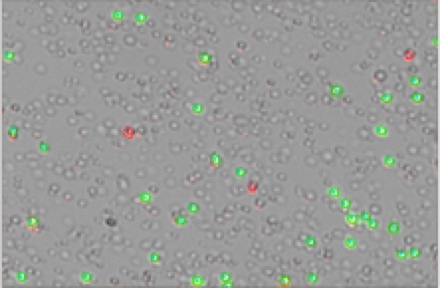
Figure 1. PBMCs Stained with AO/PI. Selective staining of live (green) and dead (red) PBMCs in the presence of RBCs (unstained).

Materials

  • CellDrop FLi Automated Cell Counter
  • Peripheral Blood Mononuclear Cells
  • DeNovix CellDrop AO/PI Assay Kit (cat #CD-AO-PI-1.5)
  • Phosphate Buffered Saline (PBS)

Procedure

Cell counting and viability measurements are performed using the DeNovix Acridine Orange (AO) / Propidium Iodide (PI) Assay. AO is a nucleic acid-binding fluorophore that is cell membrane permeable and suitable for selective staining of nucleated PBMCs. PI is a nucleic acid-binding dye that is impermeable to live cells and suitable for staining dead, nucleated PBMCs. All live, nucleated cells fluoresce green due to AO. Dead, nucleated cells are stained with both AO and PI and fluoresce red. RBCs and debris are unstained and not counted.

  1. Centrifuge PBMCs at 1000 RPM for 10 minutes, and resuspend the pellet in desired volume of PBS.
  2. Vortex cells well. Dispense desired sample volume into a microfuge tube.
  3. Mix sample with an equal volume of AO/PI reagent.
  4. Open the AO/PI app, and select the appropriate protocol. Enter any additional sample information in the Sample Name section (optional).
  5. Vortex the sample and then aspirate the sample volume indicated on the count button using a fresh pipette tip.
  6. Ensure that the arm is in the down position. Rest the pipette tip in the groove on the lower sample surface and dispense the sample into the chamber. Live images will display the sample flowing evenly across the field of view. Allow the cells to settle (~10 seconds).
  7. Focus on the cells using the green channel. Adjust exposure for the green and red fluorescent channels as necessary.
  8. Press the Count button located on the bottom right corner of the screen.

Cell Size and Fluorescence Thresholds

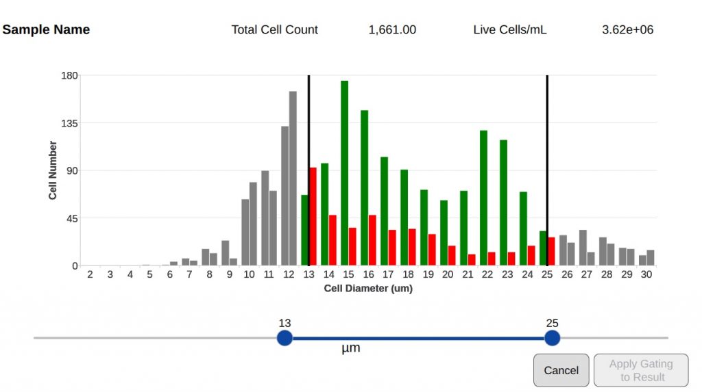
Setting an appropriate size range and fluorescence intensity for the cells of interest can exclude debris or alternative cell populations in a sample from analysis. Minimum and maximum cell diameters along with appropriate intensity thresholds can be defined and saved in protocols.

The min and max diameter can be dynamically altered using the cell size histogram once cells have been counted. The data is rapidly reanalyzed to take account of changed settings. For more advanced reanalysis, the Optimize Settings button allows the user to access and change all protocol settings on the current image and recount the sample.

Once a protocol has been optimized, the user can edit and save the original protocol with these new settings, allowing for accurate and rapid counts of future samples and standardization between different researchers.

Figure 2. Cell Size Gating. Cellular debris and non-target cell groups can be excluded from analysis based on cell diameter.

Chamber Height

The CellDrop has a unique, adjustable height sample chamber that can range from 50 to 400 µm. Setting the height at 400 µm allows for a more accurate count of low density samples.

A chamber height of 50 µm allows a higher density sample to be counted without the need for further dilution. This feature gives the CellDrop the greatest dynamic range of any image based counter on the market. The chamber height can be selected from the protocols screen. The recommended concentration range at each chamber height is detailed in Table 1.

Chamber Height
(µm)
Sample Volume
(µL)
Min Cell Density
(cells/mL)
Max Cell Density
(cells/mL)
400407.0 x 1021.0 x 105
100105.0 x 1041.0 x 107
5051.0 x 10 72.5 x 10⁷
Table 1: Chamber Height Options. 100 µm is the default chamber height and is suitable for most counts.

Summary

Automated counting of PBMC samples on the CellDrop FLi removes operator variability from the process, speeds up the workflow and enables customizable reporting and data archiving. Dual fluorescence measurements using AO/PI allow the specific identification of live and dead PBMCs in the presence of large numbers of red blood cells, platelets and cellular debris.

While AO/PI are frequently used fluorophores for this application, the CellDrop is able to measure a wide range of common fluorophores. Contact our Applications Support Team at info@denovix.com to discuss specific assay requirements.

19-NOV-2024