Introduction
The liver is one of the largest organs in the human body and plays a critical role in maintaining homeostasis through various metabolic, synthetic, and detoxifying processes. At the cellular level, hepatocytes are the principal functional units responsible for executing these complex tasks. They constitute approximately 80% of the liver mass and are pivotal in maintaining liver function and overall health.
Hepatocytes are also a widely used and a key cell type for toxicity and drug metabolism testing. Due to the sensitive nature of these types of experiments, accurate levels of hepatocyte seeding are required to obtain reproducible results.
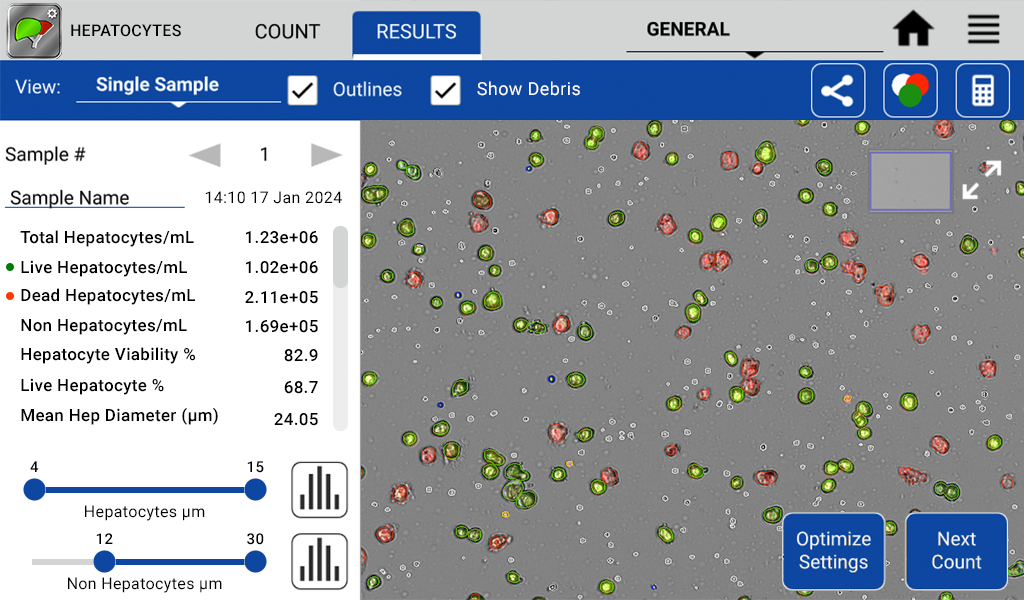
To address the needs of these researchers, DeNovix has used advanced machine learning techniques to develop algorithms to reliably report concentration and viability of hepatocyte samples. In addition, the application provides quality information such as the number of non-hepatic cells or non-cellular debris present in the specimen.
Challenges of Counting Hepatocytes Using Automated Cell Counters
Automated Cell Counters are widely used in research and offer a variety of advantages over manual counting including speed, consistent counting methods and electronic reporting and archiving of results. Hepatocytes, however, tend to be larger than most other cell types, are irregularly shaped, may have multiple nuclei per cell, and can be autofluorescent in the channels typically used for green cell counting dyes (Figure 1) due to high vitamin A content. These complexities have meant that automating the counting of this cell type has not previously been possible.
Figure 1. Left: Jurkat cells show simple round morphology and clear live (green) dead (red) staining, allowing for easy identification by a traditional cell counting algorithm. Right: hepatocytes show green autofluorescence and complex morphology.
Machine Learning Methods for Optimizing Cell Counting
To address the needs of hepatocyte researchers, DeNovix cell biologists have developed a unique application to identify and count hepatocytes. Additionally, the model was trained to identify lymphocytes, free nuclei and debris objects in the sample to give qualitative as well as quantitative information about hepatocyte isolations (Figure 2). A novel auto-thresholding feature corrects for autofluorescence and allows live / dead determination using Acridine Orange and Propidium Iodide.

Figure 2. Zoomed image of the CellDrop accurately identifying and enumerating live hepatocytes (green) dead hepatocytes (red) free nuclei (yellow outline) lymphocytes (blue outline) and erythrocytes or other debris (white).
Methods
To demonstrate the accuracy and reproducibility of the CellDrop Hepatocyte App, three species of isolated hepatocytes (canine, human and rat) were selected (BioIVT, Baltimore MD). These were thawed in accordance with the manufacturer’s instructions and resuspended in 2 mL of Invitrogro KHB buffer (BioIVT, Baltimore MD).
From this initial resuspension, 2 serial dilutions were made by adding 1 mL of this resuspension to 1 mL of KHB buffer for a total of 3 samples. The samples were then stained with AO/PI (DeNovix, Wilmington DE) and counted on a CellDrop equipped with the Hepatocyte app or stained with Trypan Blue (DeNovix, Wilmington DE) and counted on a manual hemocytometer using a brightfield microscope. Each sample was counted with 3 replicates.
Results
The CellDrop Hepatocyte App and the manual hemocytometer counts showed a very close relationship with all but one of the dilutions, showing non statistically significant differences in the counts of live hepatocytes/mL (p value < 0.02). The one dilution set that did show a significant difference was the dilution that had the lowest overall counts, which may account for this result. The degree of overlap between the two methods between species and over the range of concentrations is high and in both CellDrop and manual counts the r2 values are all greater than 0.99. Also note that the standard deviation on the CellDrop is lower than the manual hemocytometer (Figure 3).
Figure 3. Comparison of the concentration of live cell/mL for three species of hepatocytes (rat, canine and human) diluted and counted on both a CellDrop Automated Cell Counter and a manual hemocytometer. *Denotes statistically significant difference
Likewise the viability numbers between the two methods appears to be very close with no statistically significant differences between any of the nine replicates as shown in Figure 4 (p value < 0.02). Viability as well as concentration of live hepatocytes/mL did vary significantly between the three different species that were tested.
Figure 4. Comparison of the percent viability for three species of hepatocytes (rat, canine and human) diluted and counted on both a CellDrop Automated Cell Counter and a manual hemocytometer.
Discussion
The results demonstrate that the CellDrop Automated Cell Counter’s fluorescence-based Hepatocyte App gives equivalent results to a trained technician counting hepatocytes using the Trypan Blue exclusion method. While the results demonstrate the CellDrop’s high level of reproducibility, the instrument also offers several advantages over traditional methods.
First, the CellDrop has a significant time and consistency advantage compared to manual counting, especially when accounting for multiple technicians within a lab. The CellDrop also provides additional information about the sample, such as the level of debris and number of lymphocytes / free nuclei present, data which is not normally collected when counting manually. Additionally, the CellDrop stores all of the images, results and count information on board for reporting and re-analysis.
The CellDrop Hepatocyte App is a unique and highly effective tool for labs wanting to add automation to their hepatocyte workflows. For more information about the CellDrop Automated Cell Counter, click here.
Live Demo
Want to see how it works for yourself? Get a quick look at the live preview and hepatocyte count data available on the Hepatocytes app.
References
- Jin Gong et al. (2023) Hepatocytes: A key role in liver inflammation. Front Immunol. 2022; 13: 1083780.
- Lin-Gibson S, Sarkar S, Elliott J. Summary of the National Institute of Standards and Technology and US Food and Drug Administration cell counting workshop: Sharing practice in cell counting measurements, Cytotherapy 2018; 785-795
- Sarkar S, Lund S, Vyzasatya R, Vanguri P, Elliot J, Plant A, Lin-Gibson S. Evaluating the quality of a cell counting measurement process via a dilution series experimental design. Cytotherapy 2017 1509-1521
Count Cells Without Slides
If you’re interested in learning more about the CellDrop Automated Cell Counter, consider signing up for a 7-Day Free Trial, scheduling a call with our applications team, or requesting a quote. Click the buttons below to take the next step!
08-JUL-2024


