Introduction
The CellDrop Fixed Nuclei PI app provides fast, accurate counts for fixed nuclei samples using Propidium Iodide (PI), a fluorescent nuclear stain. Propidium Iodide is able to pass through the nuclear pores and intercalate between the base pairs of double stranded DNA molecules. Upon binding, the quantum yield of the dye increases by ~20-30x which makes the nuclei fluoresce red. The primary benefit of Propidium Iodide is its selective binding to nucleic acids and not to cellular debris generated during nuclei isolation workflows. This ensures that only the nuclei are counted and non-fluorescent background debris is excluded. This makes PI a more accurate dye for counting nuclei than Trypan Blue, which will also bind to cellular debris.
The CellDrop™ Automated Cell Counter paired with Propidium Iodide provides fast, accurate fixed nuclei concentration measurements, high definition images for debris assessment, and advanced sample clumping quality control metrics.

Methods for Nuclei isolation and Fixation
Generation of Single Nuclei Suspension
Fresh frozen mouse kidney was purchased from BioIVT (Baltimore, MD) and maintained at 4°C. At the time of the experiment, the mouse kidney was resuspended in nuclei separation buffer. Tissue dissociation and nuclei isolation were performed on the gentleMACS™ OctoDissociator (Miltenyi, 130-096-427) in conjunction with gentleMACS™ C tubes (Miltenyi, 130-093-237) and gentleMACS™ Octo Coolers (Miltenyi,130-130-533) using the ‘4C_nuclei_1’ protocol. Nuclei suspensions were filtered and immediately placed on ice. Initial concentration and isolation efficiency of the pre-fixed nuclei suspension was determined on a CellDrop FLi in the Nuclei AO/PI app.
Fixation of Nuclei Suspension
Fixation of the nuclei suspension was performed according to manufacturer’s guidelines from the Evercode™ Nuclei Fixation v3 (Parse, PN-NF100). Finally, an equal volume of DeNovix Propidium Iodide dye (SKU-30570) was added to an aliquoted volume of fixed nuclei suspensions and used to determine concentration and quality of the isolated nuclei suspensions in the Fixed Nuclei PI app.
The CellDrop Fixed Nuclei PI App
Accurate density determination of isolated nuclei samples is critical in all single nuclei RNA-seq (snRNA-seq) library preparation workflows, but additional information, such as degree of clumping, is typically needed when deciding whether to move forward with an experiment. Isolated nuclei suspensions with significant clumps can result in bioinformatic analysis challenges and compromise snRNA-seq dataset quality. Fixed Nuclei PI app users should refer to their nuclei isolation kit manufacturer’s guidelines for clumping or multiplet tolerance.
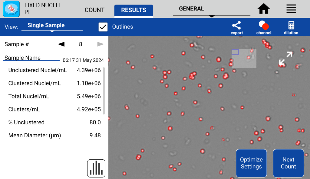
The counting algorithm in the Fixed Nuclei PI app calculates both the density of monodispersed nuclei and the density of clumped nuclei as ‘Unclustered Nuclei/mL’ and ‘Clustered Nuclei/mL.’ Additional metrics include ‘% Unclustered,’ which highlights how monodispersed the nuclei sample is prior to moving forward with library preparation. Figure 1 shows a count result of nuclei isolated from mouse kidney tissue displaying these metrics.

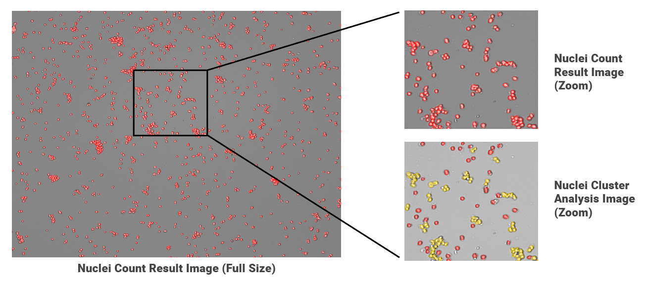
Figure 2 features a sample with multiple clumps of nuclei, classified as ‘clusters’ by the count algorithm. Further quality assessment is enabled by a suite of image export options. Users can select a ‘Cluster Image’ format (Figure 2, bottom right) that highlights individual nuclei as red and clumped nuclei as yellow with a brightfield overlay. This innovative option enables users to quickly scan images and determine the degree of clumping throughout their fixed nuclei suspensions. It also has immense utility as a sample quality check-point in single nuclei RNA-sequencing workflows. Images are automatically saved to CellDrop, creating an archive for future reference.
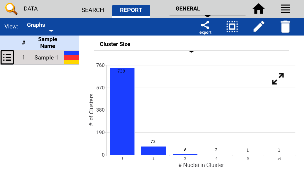
Cluster size can be graphically visualized, categorizing nuclei in up to 6 distinct cluster sizes. Figure 3 displays a results ‘Cluster Size’ graph for a sample and highlights individual nuclei, doublets and higher degree clumping. These bar graphs can be accessed in the results tab graphs view of the count app or in the Data App report tab using the graph view.

Conclusion
The CellDrop Fixed Nuclei PI app, utilizing Propidium Iodide, offers a significant advancement in the accurate and efficient quantification of fixed nuclei. By selectively staining nucleic acids and effectively excluding cellular debris, this method proves to be a more reliable alternative to traditional dyes like Trypan Blue for nuclei counting.
Beyond simple enumeration, the app provides crucial quality control metrics, specifically the differentiation and quantification of both monodispersed and clustered nuclei. The innovative ‘Cluster Image’ export option, visually highlighting nuclear clumps, coupled with the graphical representation of cluster sizes, empowers researchers with an intuitive and comprehensive understanding of their sample quality. This detailed assessment of nuclear clumping is particularly vital for sensitive downstream applications such as single nuclei RNA-sequencing, where clump-free preparations are essential for high-quality data.
The CellDrop Fixed Nuclei PI app streamlines the nuclei counting workflow while providing essential quality control features, ultimately contributing to more reliable and robust single-cell genomic analyses.
Click here to learn more about the CellDrop Automated Cell Counter or request a quote.


